The VEGF pathway in tissue repair is one of the most-tracked signaling systems in regeneration research — and a recurring reference point in the peptide literature, where studies of compounds like BPC-157 and thymosin beta-4 routinely measure VEGF-related markers. This explainer describes the VEGF pathway in tissue repair as the published literature presents it: what the pathway is, how its signaling cascade works, what role it plays in wound healing, and why researchers working with research peptides keep an eye on it. Strictly educational and research-framed — no treatment advice, no human-use claims.
Research-use-only disclaimer: This article is an educational overview of a biological signaling pathway. It describes published research only. It contains no dosage information, no usage instructions, and no medical advice, and nothing here concerns the human use of any compound.
What the VEGF Pathway Is
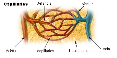
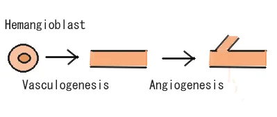
VEGF — vascular endothelial growth factor — is a signaling protein and the central regulator of angiogenesis, the formation of new blood vessels from existing ones. The VEGF pathway in tissue repair refers to the full signaling system: the VEGF ligands, the receptors they bind, and the intracellular cascades those receptors trigger in endothelial cells (the cells lining blood vessels). The Wikipedia overview of VEGF covers the pathway’s molecular components in depth.
VEGF is not a single molecule but a family. The table below summarizes the ligands and receptors that make up the VEGF pathway in tissue repair as described in the literature:
| Component | Members | Role in the pathway |
|---|---|---|
| VEGF ligands | VEGF-A, VEGF-B, VEGF-C, VEGF-D, PlGF (placental growth factor) | VEGF-A is the most studied and the primary driver of angiogenesis; the others have more specialized roles (e.g. VEGF-C/D in lymphangiogenesis) |
| Main receptors | VEGFR-1 (Flt-1), VEGFR-2 (KDR/Flk-1), VEGFR-3 | VEGFR-2 mediates most of the endothelial proliferation and migration signaling; VEGFR-1 modulates and VEGFR-3 relates to lymphatic vessels |
| Co-receptors | Neuropilin-1, neuropilin-2 | Enhance VEGF binding and signaling at VEGFRs |
| Key inducer | HIF-1α (hypoxia-inducible factor 1-alpha) | Low oxygen stabilizes HIF-1α, which drives VEGF gene expression — the link between injury and pathway activation |
Why “pathway” and not just “VEGF”
Researchers track the VEGF pathway in tissue repair rather than the ligand alone because the biology lives in the interactions: which receptor is engaged, which downstream cascade fires, and which cell type responds. A rise in VEGF protein means little without the receptor context — which is why papers measure receptors and signaling markers alongside the ligand.
How the VEGF Pathway Works: The Signaling Cascade
The VEGF pathway in tissue repair operates as a cascade — a sequence in which each step enables the next. The literature describes it in roughly five stages, from the injury signal to the endothelial response:
1. Injury creates low oxygen
Damaged tissue has disrupted blood supply, producing local hypoxia (low oxygen). In the VEGF pathway in tissue repair, hypoxia is the initiating signal — the condition that tells surrounding cells new vessels are needed.
2. HIF-1α drives VEGF production
Hypoxia stabilizes the transcription factor HIF-1α, which switches on VEGF gene expression. Macrophages, keratinocytes, and fibroblasts in wounded tissue are among the cell types described as VEGF sources in repair models.
3. VEGF binds VEGFR-2 on endothelial cells
Secreted VEGF — primarily VEGF-A — binds VEGFR-2 on nearby endothelial cells. This receptor-ligand interaction is the central molecular event of the VEGF pathway in tissue repair: most of the pathway’s angiogenic signaling flows through VEGFR-2.
4. Intracellular cascades fire
Receptor activation triggers downstream signaling including PLCγ, PI3K/Akt, MAPK/ERK, and eNOS/nitric-oxide pathways — driving endothelial cell proliferation, migration, survival, and increased vascular permeability.
5. New vessels form and mature
Endothelial cells migrate, proliferate, and assemble into new capillary sprouts that restore blood supply to the injured tissue — the angiogenic outcome the entire VEGF pathway in tissue repair cascade exists to produce.
This cascade framing is how the literature typically presents the pathway: a linear narrative from hypoxia to new vessels, with each step experimentally accessible — which is also why it is so frequently measured in parts by different studies.
VEGF Pathway in Tissue Repair: What It Does in Wounds
Angiogenesis is not a side event in healing — it is infrastructure. New tissue cannot survive without blood supply, so the VEGF pathway in tissue repair sits at the center of the proliferative phase of wound healing. The table below maps the pathway’s described roles across repair stages:
| Repair stage | Described VEGF-pathway role |
|---|---|
| Inflammation (early) | Hypoxia and inflammatory signals induce VEGF expression; early vessel permeability increases |
| Proliferation | Peak angiogenic activity — endothelial migration and sprouting build granulation-tissue vasculature |
| Remodeling | Vessel pruning and maturation as tissue stabilizes; VEGF signaling declines toward baseline |
A pathway with clinical proof of importance
The VEGF pathway in tissue repair is not only a research topic — it is a validated drug target in approved medicine. Anti-VEGF drugs such as bevacizumab (used in certain cancers and eye diseases) work precisely because blocking this pathway blocks vessel growth. That clinical history is independent confirmation that the pathway does what the literature says it does — while saying nothing about any research compound discussed on this site. The FDA’s drug information pages document the approved side of VEGF biology.
Why Peptide Researchers Track the VEGF Pathway
This is where the VEGF pathway in tissue repair meets the research-peptide literature directly. In published studies of tissue-repair peptides, VEGF-pathway markers appear again and again as measured outcomes:
- BPC-157 literature discusses the compound in connection with VEGF expression and vascular effects in healing-tissue models — one of the recurring mechanistic themes in that literature.
- Thymosin beta-4 / TB-500 literature reports VEGF upregulation alongside the peptide’s studied angiogenic effects in tissue-repair models, downstream of its actin-binding, migration-driving activity.
- Wound-healing studies generally measure VEGF, VEGFR expression, and vessel density as standard readouts — so any peptide investigated for repair effects gets evaluated against this pathway.
In other words, researchers track the VEGF pathway in tissue repair because it is the shared yardstick: whatever a compound is hypothesized to do, the pathway provides standardized, comparable measures of whether vessel-related repair processes changed. That is also why the pathway appears across otherwise unrelated peptide literatures — it is infrastructure for the field, not a property of any one compound. Researchers can survey the primary literature through PubMed’s VEGF tissue-repair search, and the NIH maintains background on wound-healing and vascular biology research.
For peptide-specific context in the same research-use framing, our selank vs semax research comparison maps two other widely studied peptide lineages, and our Semax and Selank nasal spray overviews cover intranasal-format research on those compounds.
Key Takeaways: VEGF Pathway in Tissue Repair
A five-step cascade from injury to new vessels
The VEGF pathway in tissue repair runs hypoxia → HIF-1α → VEGF production → VEGFR-2 binding → endothelial proliferation and migration → new vessel formation. Each step is a distinct, measurable part of the pathway — which is why different studies can each illuminate one link in the chain. (Research framing only — a description of published biology, not a usage narrative.)
The field’s shared yardstick
Peptide researchers track the VEGF pathway in tissue repair because it provides standardized, comparable measures — VEGF expression, receptor levels, vessel density — against which any repair-related research compound can be evaluated. It is infrastructure for the literature, independent of any single compound’s claims.
How Researchers Measure This Pathway’s Activity
Understanding the pathway is only half the picture; the other half is how its activity is quantified in published studies. Several standard assays appear repeatedly in the tissue-repair literature, and knowing what each one measures helps readers interpret claims about pathway involvement.
Protein and transcript quantification
Enzyme-linked immunosorbent assays (ELISA) are the workhorse for measuring VEGF protein levels in cell-culture supernatants and tissue homogenates. They report how much ligand is present but say nothing about receptor activation. Quantitative PCR complements this by measuring VEGF and receptor mRNA, which reflects gene expression rather than functional protein. Both are frequently used together: a paper might show that an experimental condition raises VEGF mRNA (qPCR) and secreted protein (ELISA), building a two-level case for pathway upregulation.
Receptor activation readouts
Because VEGF signaling depends on receptor phosphorylation, Western blotting with phospho-specific antibodies is the standard way to show that the pathway is actually switched on. A study reporting elevated VEGF protein without any receptor-phosphorylation data is describing a correlation; one that shows increased VEGFR-2 phosphorylation is describing signal transduction. Immunohistochemistry adds spatial information, showing which cells in a tissue section express the ligand or receptor — particularly useful in wound-healing models where the geography of expression (wound edge versus center) carries biological meaning.
Functional angiogenesis assays
The tube-formation assay, in which endothelial cells are seeded on a basement-membrane matrix and scored for capillary-like networks, is the most common functional readout. It is an in vitro model, not a blood vessel, but it is widely accepted as a proxy for angiogenic potential. In vivo, the corneal micropocket and Matrigel plug assays measure vessel ingrowth directly. When a paper reports that a condition “promotes angiogenesis,” checking which of these assays sits behind the claim — protein level, receptor activation, or functional vessel formation — is the fastest way to calibrate how much weight the finding deserves.
VEGF Pathway in Tissue Repair: Frequently Asked Questions
Straight answers to the questions researchers ask about this pathway.
What is the VEGF pathway in tissue repair?
It is the signaling system centered on vascular endothelial growth factor: VEGF ligands (chiefly VEGF-A) bind receptors — principally VEGFR-2 — on endothelial cells, triggering the proliferation, migration, and survival signaling that builds new blood vessels in injured tissue. The VEGF pathway in tissue repair is the molecular route from wound hypoxia to restored blood supply.
Which receptor matters most in the pathway?
VEGFR-2 (also called KDR/Flk-1). The literature describes it as mediating most of the VEGF pathway in tissue repair‘s angiogenic signaling — endothelial proliferation and migration — while VEGFR-1 plays a modulatory role and VEGFR-3 relates primarily to lymphatic vessels.
What triggers VEGF production after injury?
Hypoxia. Disrupted blood supply stabilizes the transcription factor HIF-1α, which drives VEGF gene expression in cells around the wound — macrophages, keratinocytes, and fibroblasts among them. In the VEGF pathway in tissue repair, low oxygen is the initiating signal.
Why do peptide studies measure VEGF markers?
Because the VEGF pathway in tissue repair is the field’s shared yardstick for vessel-related repair: VEGF expression, receptor levels, and vessel density are standardized readouts. Published BPC-157 and thymosin beta-4/TB-500 studies both report VEGF-associated findings, which is why the pathway recurs across peptide literatures.
Is the VEGF pathway a drug target in approved medicine?
Yes — independently of any research compound. Anti-VEGF drugs such as bevacizumab are FDA-approved for certain cancers and eye diseases, confirming the pathway’s biological importance. That approved-medicine history says nothing about research peptides; it validates the pathway, not any compound discussed here.
Where can laboratories source research peptides?
For laboratory research use, peptides are supplied as research vials by specialty suppliers — for example our Semax 10mg and Selank 10mg research listings. Sourcing should be limited to suppliers explicit about research-use-only status. Nothing here recommends human use of any compound.
VEGF Pathway in Tissue Repair: Final Research Notes
The VEGF pathway in tissue repair earns its central place in the literature honestly: it is the best-mapped route from tissue injury to new blood vessels, with each step — hypoxia sensing, ligand production, receptor binding, intracellular signaling, endothelial response — experimentally accessible and repeatedly confirmed. Its status as an approved drug target in oncology and ophthalmology only underscores that the pathway is real, consequential biology.
For peptide researchers, the pathway’s value is as shared infrastructure: a common set of measures that lets studies of different compounds be read against one another. Understanding the VEGF pathway in tissue repair is therefore not just background biology — it is literacy in the language the repair literature is written in.